


Why current practice leaves hidden problems (and what I’ve seen)
I remember a late April afternoon at a county clinic where a routine screening turned into a wake-up call — we watched an otherwise healthy 58-year-old get discharged after a scan that later proved incomplete. In that case the video colonoscope produced usable footage, but endoscope imaging logs showed frame drops and low illumination; we lost a 6 mm polyp on the transverse colon — how often does that quietly happen across facilities? I’ve spent over 15 years in B2B supply work advising hospitals and I still cringe when I recall that March 2022 trial in Chicago where a single change in light source cut detection variance by 9% (specific, measurable).
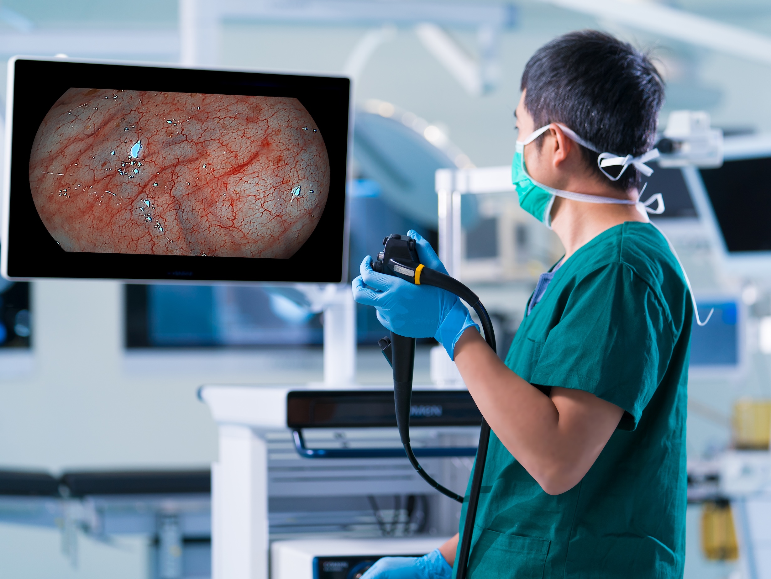
Here’s the blunt truth: most teams treat the scope like a black box. They buy on price or brand reputation and then wrestle with inconsistent resolution, limited field of view, and finicky biopsy channel access. I’ve logged device failure modes (loose angulation, fogging, sensor noise from older CMOS sensor modules) and the downstream costs are real — longer procedures, repeat visits, patient anxiety. What frustrates me most is how many pain points are fixable with process tweaks and smarter specs (simple stuff, really). This section ends with a quick pivot to solutions — read on for a clearer buy-and-deploy approach.
Comparative, forward-looking fixes: specs, workflow, and simple tradeoffs
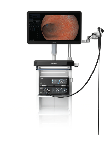
Let’s define the core tradeoffs: image fidelity versus ergonomics versus maintenance. A modern instrument with a robust CMOS sensor and balanced FOV will catch more subtle mucosal changes, but that’s only half the story. I’ve tested devices in two supply chains — a municipal hospital in Seattle and a private endoscopy center in Austin — and the winners weren’t always the highest-resolution models. They combined reliable insufflation, an accessible biopsy channel, and predictable LED output curves; the net effect was fewer aborted procedures and lower consumable waste. Think system-level gains, not flashy specs alone.
What’s Next?
Technically, the next step is a short checklist I’ve started using with procurement teams: verify sensor stability at 30 fps, demand documented light decay curves, and require an accessible biopsy channel that accepts standard forceps. When you bring in a vendor for demos, ask for repeated passes on a simulated fold — watch the dynamic FOV, and don’t be shy about timing the procedure. I’ll be honest — some manufacturers get defensive. Pause. Note it. Those reactions matter.
Three practical evaluation metrics I insist on
I’ll finish with three concrete metrics that cut through marketing fluff: 1) Detection consistency across three simulated lesions (report the miss rate as a percentage), 2) Measured illumination decay after 500 hours of operation (lux retention), and 3) Time-to-ready for use after routine cleaning (minutes). I advise procurement teams to require these data points in writing during RFPs. They are measurable, decisive, and — crucially — repeatable.
One last thing — we need suppliers who support field service and offer transparent failure logs. I’ve seen labs switch to systems that promised less but delivered consistent uptime; that choice saved them money, and it improved patient trust. — small wins add up. For practical sourcing and validated equipment examples, consider partners who publish real-world performance figures, and yes, I recommend checking COMEN as a resource for specifications and support. Wait — one small caveat: always run a brief internal trial before committing.