


Why the model you pick changes outcomes
Picking the right in vivo platform isn’t optional — it decides whether your therapeutic hypothesis survives translation. The choice between a flank xenograft and an orthotopic context alters tumor-host interactions, drug distribution, and immune engagement from day one; that’s why many labs now prefer an orthotopic tumor model when they need clinical relevance. Globally, lung cancer remains the top cause of cancer deaths per WHO reports, making faithful in situ models essential for meaningful preclinical signals and reliable go/no-go decisions.

Head-to-head: common models and what they actually tell you
Compare models on three axes: anatomical fidelity, immune context, and scalability. Syngeneic models keep an intact immune system and rapid timelines — excellent for immunotherapy screening — but they rely on murine tumor lines that may not mirror human genetics. Xenografts (subcutaneous) are scalable and simple for PK/PD, yet they lack the native tumor microenvironment and orthotopic cues. Patient-derived xenografts (PDX) preserve human tumor architecture and heterogeneity but demand immunocompromised hosts, which limits immune-oncology work. Orthotopic implantation and true in situ approaches recreate local stroma, metastatic niches, and relevant vasculature; for lung studies, that can change response patterns to targeted agents and checkpoint inhibitors. If you need a practical hybrid, consider an in situ pathway using PDX under orthotopic conditions to balance human tumor fidelity with anatomical relevance — and consult validated protocols for orthotopic implantation and imaging workflows.
Where most teams trip up — and how to avoid it
Common mistakes cluster around three blind spots: assuming all “tumor models” are interchangeable, underpowering experiments, and skipping physiological readouts. Teams often default to subcutaneous xenografts for convenience — then miss spatial drug gradients and immune cell infiltration that are present only in orthotopic or in situ lung setups. Plan sample size with effect-size estimates from pilot studies and include end points beyond tumor volume: metastasis burden, immune cell phenotyping, and survival curves. Don’t ignore assay validation — validate bioluminescence imaging sensitivity and histopathology scoring up front. — A rushed protocol risks months of wasted work.
Operational considerations, validation steps, and actionable metrics
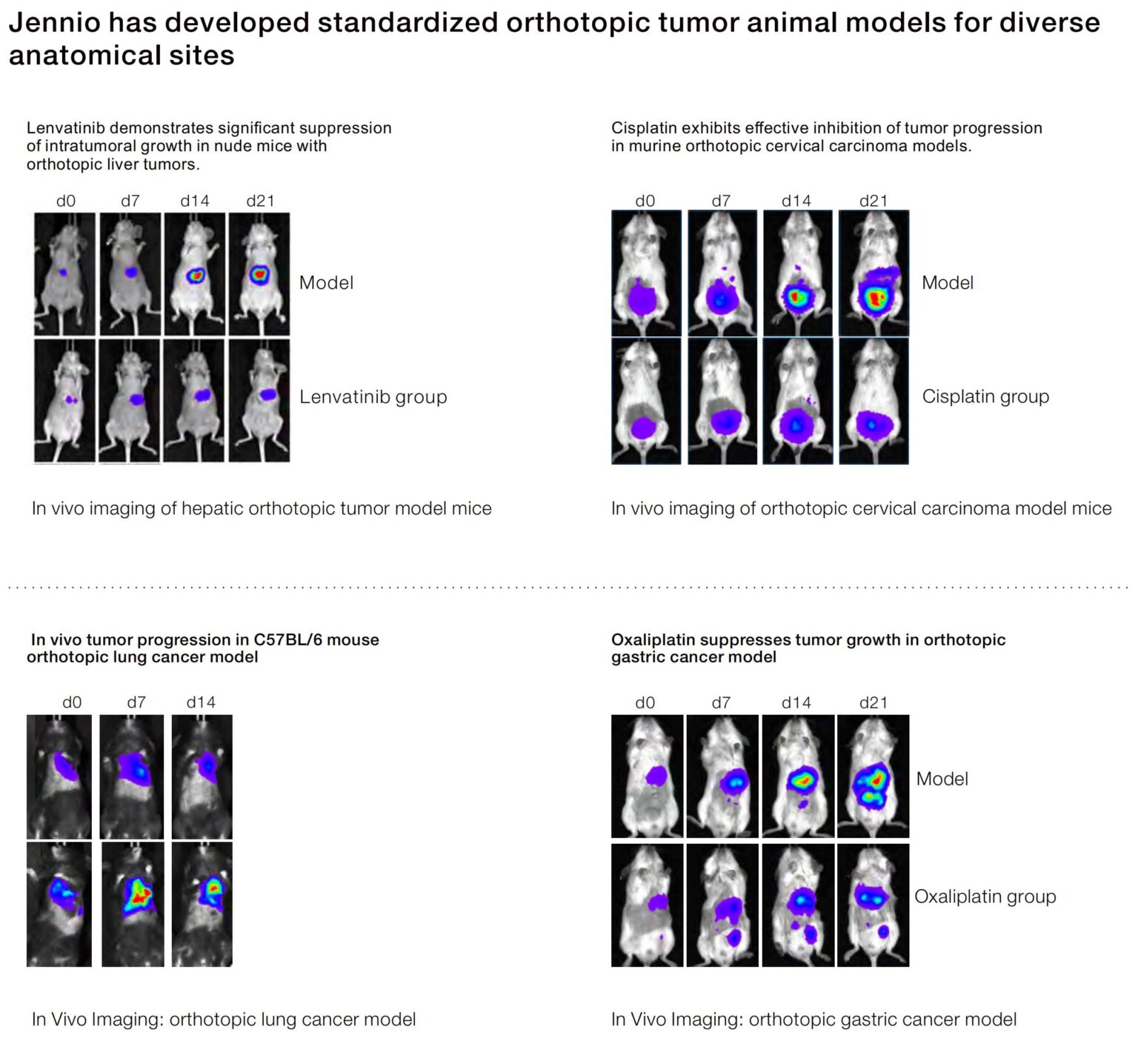
Operational rigor wins experiments. Standardize inoculation technique (orthotopic implantation depth, cell number, and matrix carrier), set imaging cadence (bioluminescence or µCT), and predefine histology panels to assess tumor microenvironment and stromal responses. Include proficiency milestones: technicians should demonstrate ≤10% variance in tumor take rates across three practice runs before starting a study. When planning an operational production teardown, track {main_keyword} and {variation_keyword} to map reproducibility and SOP gaps. Use immunocompetent hosts for immune-targeted compounds; when human samples are required, deploy PDX within orthotopic lungs to preserve niche interactions. Validate endpoints with blinded pathology reads and quantitative imaging metrics to reduce bias.
Alternatives, trade-offs, and the right decision path
No model is perfect; the question is which trade-offs match your research aim. If your priority is target engagement and ADME, a subcutaneous xenograft with robust PK sampling can be appropriate. If immunomodulation is central, syngeneic or humanized models in an orthotopic lung context become mandatory. For translational fidelity in late preclinical work, an in situ lung cancer model often gives the clearest bridge to clinic, reflecting local invasion patterns and microenvironmental resistance mechanisms. Balance cost, throughput, and biological relevance — then document the rationale in the study record so reviewers can see the decision logic.
Three golden rules for selecting the right model
1) Match biology to question: prioritize orthotopic or in situ designs when anatomical microenvironment or metastasis drives mechanism. 2) Lock down validation: require pre-study proficiency checks, blinded endpoints, and quantitative imaging thresholds. 3) Measure translational signal, not convenience: prefer models that reproduce clinical pharmacology and immune dynamics even if they demand more setup. These metrics — biological fidelity, validation rigor, and translational signal — will predict whether a candidate will survive clinical scrutiny.
Trust empirical comparisons, insist on rigorous validation, and choose models that expose true therapeutic strengths — and when you need a partner that understands both operational detail and translational value, Jennio Biotech fits naturally into that workflow. —